
Mouse Secondary Antibodies

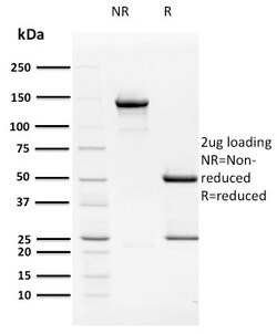
Biotium HPV16 E2 (Human Papilloma Virus 16)(TVG 261), CF488A conjugate, 0.1mg/mL
The human papilloma virus (HPV) family of DNA tumor viruses includes HPV16, a strain that is responsible for the largest number of cases of cervical cancers linked to the family. HPV16E1 and HPV16E2 are proteins that are involved in the regulation of viral DNA replication and are important for infected cell homeostasis. HPV16E2 specifically regulates the expression of the E6 and E7 oncoproteins by binding to four sites within the viral long control region, possibly involving interactions with nuclear hormone receptors. Integration of the HPV genome into the host DNA usually disrupts the HPV16E2 gene open reading frames, resulting in an overexpression of E6 and E7 genes, an event that may lead to the malignant transformation of cervical cancer. HPV16E2 is also able to induce apoptotic cell death via two pathways: the first through the binding of p53 and the second through the binding of the viral genome. Primary antibodies are available purified, or with a selection of fluores

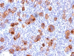
Biotium CD30 / TNFRSF8 (Hodgkin & Reed-Sternberg Cell Marker) (rKi-1/779), CF488A conjugate, 0.1mg/mL
This antibody recognizes a single chain glycoprotein of 105/120 kDa, identified as CD30/Ki-1. CD30 is synthesized as a 90 kDa precursor, which is processed in the Golgi complex into a membrane-bound phosphorylated mature 105/120 kDa glycoprotein. In Hodgkin's disease, CD30/Ki-1 antigen is expressed by mononuclear-Hodgkin and multinucleated Reed-Sternberg cells. It is also expressed by the tumor cells of a majority of anaplastic large cell lymphomas as well as by a varying proportion of activated T and B cells. This MAb distinguishes large cell lymphomas derived from activated lymphoid cells from histiocytic malignancies and lymphomas derived from resting and precursor lymphoid cells or from anaplastic carcinomas. About one third of the Ki-1 positive lymphomas lack the leukocyte common antigen (CD45).Primary antibodies are available purified, or with a selection of fluorescent CF Dyes and other labels. CF Dyes offer exceptional brightness and photostability. Note: Conjugate
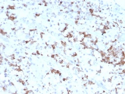
Biotium Prolactin (Pituitary Tumor Marker)(PRL/2641), 1mg/mL
Prolactin is a growth factor that is secreted by the anterior pituitary. It is necessary for the proliferation and differentiation of the mammary glands. Prolactin is useful in the classification of pituitary tumors and study of pituitary disease. It also plays a role in the development of mammary cancer, functioning dually as a mitogen and a differentiating agent. Primary antibodies are available purified, or with a selection of fluorescent CF Dyes and other labels. CF Dyes offer exceptional brightness and photostability. Note: Conjugates of blue fluorescent dyes like CF405S and CF405M are not recommended for detecting low abundance targets, because blue dyes have lower fluorescence and can give higher non-specific background than other dye colors.
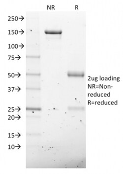
Biotium NKX2.8(NKX28/2547), CF568 conjugate, 0.1mg/mL
The protein encoded by this gene is a homeobox-containing developmental regulator associated with liver development. The encoded protein binds to the alpha-fetoprotein (AFP) gene promoter and increases the expression of AFP. This gene is overexpressed in some lung cancers and is linked to poor patient survival, possibly due to its resistance to cisplatin. This gene is aberrantly methylated in pancreatic cancer, deleted in squamous cell lung carcinomas, and acts as a tumor suppressor in esophageal cancer. Mutations in this gene may also be a cause of neural tube defects. Primary antibodies are available purified, or with a selection of fluorescent CF Dyes and other labels. CF Dyes offer exceptional brightness and photostability. Note: Conjugates of blue fluorescent dyes like CF405S and CF405M are not recommended for detecting low abundance targets, because blue dyes have lower fluorescence and can give higher non-specific background than other dye colors.

Biotium Calretinin / Calbindin 2 (Mesothelioma Marker)(CALB2/2603), CF568 conjugate, 0.1mg/mL
Calretinin (also known as Calbindin 2) is a member of the troponin C superfamily. Proteins in this family have 6 EF-hand domains which bind to calcium. Calretinin is a vitamin D-dependent calcium-binding protein involved in calcium signaling. It is present in subsets of neurons throughout the brain and spinal chord, including sensory ganglia. Antibodies to calretinin are useful in differentiating mesothelioma from adenocarcinomas of the lung. They can also aid in differentiating adrenal cortical neoplasms from pheochromocytomas. Primary antibodies are available purified, or with a selection of fluorescent CF Dyes and other labels. CF Dyes offer exceptional brightness and photostability. Note: Conjugates of blue fluorescent dyes like CF405S and CF405M are not recommended for detecting low abundance targets, because blue dyes have lower fluorescence and can give higher non-specific background than other dye colors.
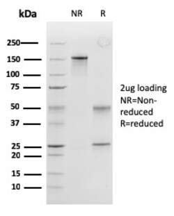
Biotium Peroxiredoxin 4 (Prognostic Marker for Lung SqCC)(CPTC-PRDX4-2), CF740 conjugate, 0.1mg/mL
The peroxiredoxin (PRX) family comprises six antioxidant proteins, PRX I, II, III, IV, V and VI, which protect cells from reactive oxygen species (ROS) by preventing the metal-catalyzed oxidation of enzymes. The PRX proteins primarily utilize thioredoxin as the electron donor for antioxidation, although they are fairly promiscuous with regard to the hydroperoxide substrate. In addition to protection from ROS, peroxiredoxins are also involved in cell proliferation, differentiation and gene expression. PRX I, II, IV and VI show diffuse cytoplasmic localization, while PRX III and V exhibit distinct mitochondrial localization. The human PRX IV gene is expressed in many tissues. It exists as a precursor protein, which is only detected in testis, and aprocessed secreted form. PRX IV is highly expressed in lung cancer and is necessary for the promotion of lung cancer in vitro. Studies have demonstrated that PRX IV positive expression is significantly correlated with recurrences and

Biotium AKT1 (Prognostic Marker for Neuroendocrine Tumors)(AKT1/2491), Biotin conjugate, 0.1mg/mL
AKT1 is a 62 kDa protein and member of the serine/threonine kinase AKT family. AKT1 is also known as Protein Kinase B (PKB) or Rac Protein Kinase Alpha. The Akt family also includes AKT2. Activation of AKT1 by insulin or insulin-growth factor-1 (IGF-1) results in phosphorylation of both Thr 308 and Ser 473. AKT proteins become phosphorylated and activated in insulin/IGF-1-stimulated cells by an upstream kinase(s), and the activation of AKT1 and AKT2 is inhibited by the PI kinase inhibitor wortmannin. Primary antibodies are available purified, or with a selection of fluorescent CF Dyes and other labels. CF Dyes offer exceptional brightness and photostability. Note: Conjugates of blue fluorescent dyes like CF405S and CF405M are not recommended for detecting low abundance targets, because blue dyes have lower fluorescence and can give higher non-specific background than other dye colors.
Biotium RCAS1 / Estrogen Receptor Binding Site Associated, Antigen 9(CPTC-EBAG9-2), CF568 conjugate, 0.1mg/mL
EBAG9, also known as RCAS1, is an estrogen-transcribed protein. Soluble and membranous RCAS1 proteins may play a role in the immune escape of tumor cells by promoting T lymphocyte inhibition of growth and apoptosis. RCAS1 is expressed in a wide variety of cancers, including uterine, ovarian, and lung cancer cells, and acts as a ligand for a putative receptor present on peripheral lymphocytes. RCAS1 is highly expressed not only in cancer cells but also in non-tumor bile duct cells subject to immune attack. RCAS1 inhibits the in vitro growth of receptor-expressing cells and induces apoptosis, contributing to the ability of tumor cells to evade host immune surveillance. High expression of RCAS1 significantly correlates with tumor progression and with poor outcome for many cancer patients. Primary antibodies are available purified, or with a selection of fluorescent CF Dyes and other labels. CF Dyes offer exceptional brightness and photostability. Note: Conjugates of blue fluo
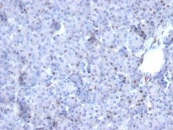
Biotium AKT1 (Prognostic Marker for Neuroendocrine Tumors)(AKT1/2784), CF740 conjugate, 0.1mg/mL
This antibody recognizes a protein of 62 kDa, which is identified as AKT1. The serine/threonine kinase Akt family contains several members, including Akt1 (also designated PKB or RacPK), Akt2 (also designated PKBbeta or RacPK-beta) and Akt3 (also designated PKB-gamma or thymoma viral proto-oncogene 3), which exhibit sequence homology with the protein kinase A and C families and are encoded by the c-Akt proto-oncogene. All members of the Akt family have a Pleckstrin homology domain. Akt1 and Akt2 are activated by PDGF stimulation. This activation is dependent on PDGFR-beta tyrosine residues 740 and 751, which bind the subunit of the phosphatidylinositol 3-kinase (PI 3-kinase) complex. Activation of Akt1 by insulin or insulin-growth factor-1 (IGF-1) results in phosphorylation of both Thr 308 and Ser 473. Akt proteins become phosphorylated and activated in insulin/IGF-1-stimulated cells by an upstream kinase(s), and the activation of Akt1 and Akt2 is inhibited by the PI kinase i
Biotium HER-4 / ERBB4(ERBB4/2581), Biotin conjugate, 0.1mg/mL
The EGF receptor family comprises several related receptor tyrosine kinases that are frequently overexpressed in a variety of carcinomas. Members of this receptor family include EGFR (HER1), Neu (ErbB-2, HER2), ErbB-3 (HER3) and ErbB-4 (HER4), which form either homodimers or heterodimers upon ligand binding. The gene encoding ErbB-4 is expressed as a full-length protein, which produces a short membrane-anchored cytoplasmic domain fragment and a long ectodomain fragment. The short fragment is heavily tyrosine phosphorylated and possesses tyrosine kinase catalytic activity toward an exogenous substrate. Proteolytic cleavage of ErbB-4 is promoted by the binding of heregulin. ErbB-4 is involved in cell proliferation and differentiation and its expression is highest in breast carcinoma cell lines, normal skeletal muscle, heart, pituitary, brain and cerebellum. Its expression in breast cancer, pediatric brain cancer and other types of carcinomas has been reported in studies which s
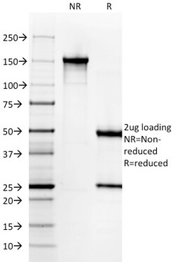
Biotium Gastrin(GAST/2631), Biotin conjugate, 0.1mg/mL
Gastrin, is a hormone that normally formed by mucosal cells in the gastric antrum and by the D cells of the pancreatic islets. Its primary function is to stimulate secretion of HCl by the gastric mucosa. HCl, in turn, inhibits gastrin formation. It also responsible for stimulating smooth muscle contraction and increasing blood circulation and water secretion in the stomach and intestine. Gastrin is regulated by epidermal growth factor in both mice and humans. Gastrin is excreted in excess by pancreatic tumors in the Zollinger-Ellison syndrome. Gastrin-Releasing Peptide (GRP) stimulates the release of gastrin as well as other gastrointestinal hormones and also acts as an autocrine growth factor for certain cell types. High levels of GRP are found in the human lung just after birth and levels decrease thereafter in parallel with the observed disease in a number of pulmonary neuroendocrine cells. GRP is known to promote lung tumorigenesis in model systems. Primary antibodies are
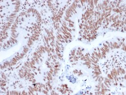
Biotium NRF1(NRF1/2608), CF740 conjugate, 0.1mg/mL
The NF-E2 DNA binding protein is composed of two subunits, p45 and MafK, and it regulates expression of globin genes in developing erythroid cells through interaction with Maf recognition elements (MAREs). A family of NF-E2 related proteins, which are collectively known as the Cap 'n' collar (CNC) family and include Nrf1 (also designated TCF11), Nrf2 and Nrf3, are bZIP transcription factors that heterodimerize with Maf proteins to bind MARE sequences. The Nrf proteins also bind the antioxidant response element (ARE) and are implicated in the regulation of detoxification enzymes and the oxidative stress response. They do so by heterodimerizing with Jun family members (c-Jun, JunB and JunD) to activate gene expression, specifically the detoxifying enzyme, NQO1. Nrf2 is widely expressed and is thought to translocate to the nucleus after treatment with xenobiotics and antioxidants, which stimulate its release from a repressor protein Keap1. Nrf3 is highly expressed in placenta, B
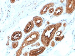
Biotium HER-4 / ERBB4(ERBB4/2581), CF568 conjugate, 0.1mg/mL
The EGF receptor family comprises several related receptor tyrosine kinases that are frequently overexpressed in a variety of carcinomas. Members of this receptor family include EGFR (HER1), Neu (ErbB-2, HER2), ErbB-3 (HER3) and ErbB-4 (HER4), which form either homodimers or heterodimers upon ligand binding. The gene encoding ErbB-4 is expressed as a full-length protein, which produces a short membrane-anchored cytoplasmic domain fragment and a long ectodomain fragment. The short fragment is heavily tyrosine phosphorylated and possesses tyrosine kinase catalytic activity toward an exogenous substrate. Proteolytic cleavage of ErbB-4 is promoted by the binding of heregulin. ErbB-4 is involved in cell proliferation and differentiation and its expression is highest in breast carcinoma cell lines, normal skeletal muscle, heart, pituitary, brain and cerebellum. Its expression in breast cancer, pediatric brain cancer and other types of carcinomas has been reported in studies which s
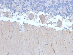
Biotium ATG5 (Autophagy Marker)(ATG5/2492), CF568 conjugate, 0.1mg/mL
Autophagy protein 5 (ATG5), in combination with autophagy protein 12, functions as an E1-like activating enzyme in a ubiquitin-like conjugating system. ATG5 is involved in several cellular processes, including autophagic vesicle formation, mitochondrial quality control after oxidative damage, negative regulation of the innate antiviral immune response, lymphocyte development and proliferation, MHC II antigen presentation, adipocyte differentiation, and apoptosis. The ATG5 protein is essential for autophagy; a process that is usually beneficial for cells to self-degrade their own components when they are no longer useful. Primary antibodies are available purified, or with a selection of fluorescent CF Dyes and other labels. CF Dyes offer exceptional brightness and photostability. Note: Conjugates of blue fluorescent dyes like CF405S and CF405M are not recommended for detecting low abundance targets, because blue dyes have lower fluorescence and can give higher non-specifi
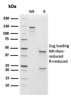
Biotium Human IgM (Immunoglobulin Mu Heavy Chain) (B-Cell Marker)(rIGHM/1623), Biotin conjugate, 0.1mg/mL
Recognizes a protein of 75 kDa, identified as mu heavy chain of human immunoglobulins. It does not cross-react with alpha (IgA), gamma (IgG), epsilon (IgE), or delta (IgD), heavy chains, T-cells, monocytes, granulocytes, or erythrocytes. Monomeric IgM is expressed as a membrane bound antibody on the surface of B cells and as a pentamer when secreted by plasma cells. IgM antibody is prominent in early immune responses to most antigens. Aberrant levels are associated with immune deficiency states, hereditary deficiencies, myeloma, Waldenstrom's macroglobulinemia, chronic infection and hepatocellular disease. This MAb is useful in the identification of leukemias, plasmacytomas, and certain non-Hodgkin's lymphomas. The most common feature of these malignancies is the restricted expression of a single heavy chain class. Demonstration of clonality in lymphoid infiltrates indicates that the infiltrate is clonal and therefore malignant. Primary antibodies are available purified, or w